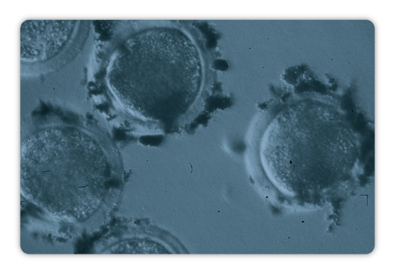
In the last decades, assisted reproductive technologies have undergone a rapid development, mainly due to the increase in fertility problems in human couples. Moreover, in the animal world, these technologies are also becoming important tools, both to increase the efficiency and quality of livestock production and to help with the conservation of wild endangered species. However, one of the major limitations in reproductive technologies is derived from the manipulation of oocytes and embryos, since they require conditions of maximum control to preserve their fertilizing ability in the case of the oocyte and development quality in the case of the embryos. Processes such as in vitro maturation of oocytes, in vitro fertilization, embryonic culture and development or vitrification require the manipulation of oocytes and embryos, both to displace them to provide required media and reagents or to immobilize them in supports for better visualization and easy handle for transfer.
In this scenario, NanoRep emerges as a new assisted reproductive methodology that will allow to manipulate oocytes and embryos without physical contact. Our specially designed nanoparticles use a novel molecular method to join with the natural glycoprotein matrix that surrounds oocytes and embryos, the zona pellucida. Once our nanoparticles are attached to them, oocyte and embryos can be moved towards any desired direction or stay fixed avoiding unnecessary displacements without physical manipulation, but by applying controlled magnetic fields. This new technology makes oocytes and embryos handling easier than ever, opening up a whole new range of applications to improve assisted reproduction lab workflows and protocols.
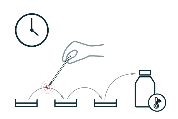
Improve the efficiency of the vitrification process by a quicker “pick up process” of oocytes or embryos using a magnetic device and avoiding loss of valuable genetic material.
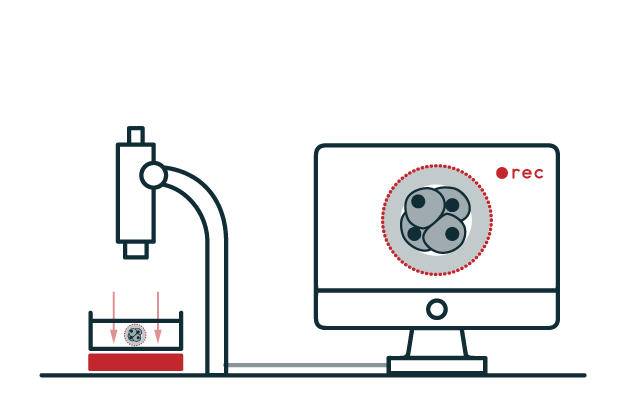
Improve the tracking and record of embryos development by fixing them to a surface, avoiding thus undesired movements without the need of using plates with sophisticated surfaces.
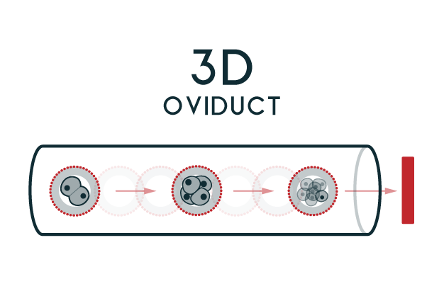
Recreate the movement of the oocyte or early embryo stage through a 3D duct, to mimic what happens naturally in the oviduct, being the in vitro conditions closer to how it occurs in vivo.
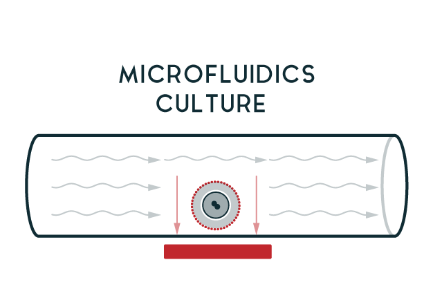
Allow the attachment of the oocyte and the embryo to a fixed point, and thus favor the culture of these cells in an environment where flows are generated to improve the quality of their development.

Biochemistry degree (graduated in 2000) and European PhD in 2006 (University of Murcia) working on the characterization of the matrix that surrounds the egg and embryo. During her postdoc, Maria worked with Dr. Jurrien Dean at National Institute of Health, Bethesda, MD, USA (2006-2010) where she applied molecular techniques to study gametes recognition. She has contributed with numerous scientific manuscripts, chapters’ books and international conferences. Maria has developed research projects as IP focused in the fertilization event, describing both the molecules responsible for the recognition of gametes, as the oviduct proteins involved in the process of fertilization and embryonic development. His research during the last years consists in applying molecular biology techniques based on the use of micro and nanospheres in the development of new techniques applied to reproduction.

Veterinary degree (graduated in 2002) and European PhD with the extraordinary award in 2007 (University of Murcia) working on the production of porcine transgenic animals. More than 15 years’ experience in teaching and research. He has developed research activities at University of Baja California (México), University of Okayama (Japan), University of Bologna and Pisa (Italy), University of Michigan State (USA) and University of Massachusetts (USA). He has contributed with numerous scientific manuscripts and chapters of books and has obtained different research awards. Author of several patents. His research is focused mainly on artificial insemination, sperm selection within the uterus, nanobiotechnology, sperm-uterus interaction, and sperm capacitation signal pathways.
Jiménez-Movilla M, Hamze JG, Romar R. Oolemma Receptors in Mammalian Molecular Fertilization: Function and New Methods of Study. Front Cell Dev Biol. 2021 May 19; 9:662032. doi.org/10.3389/fcell.2021.662032
Moros-Nicolás C, Chevret P, Jiménez-Movilla M, Algarra B, Cots-Rodríguez P, González-Brusi L,Avilés M, Izquierdo-Rico MJ. New Insights into the Mammalian Egg Zona Pellucida. Int J Mol Sci. 2021 Mar 23; 22(6):3276. doi.org/10.3390/ijms22063276
Leopoldo González-Brusi, Blanca Algarra, Carla Moros-Nicolás, Maria José Izquierdo-Rico, Manuel Avilés *, Maria Jiménez-Movilla *. A Comparative View on the Oviductal Environment during the Periconception Period. Biomolecules. 2020 Dec 17;10(12):1690. doi.org/10.3390/biom10121690
Julieta Gabriela Hamze, Maria Jimenez-Movilla*, Raquel Romar*. Sperm-Binding Assay Using an In Vitro 3D Model of the Mammalian Cumulus-Oocyte Complex. Curr Protoc Toxicol. 2020 Dec. 86(1):e100. doi.org/10.1002/cptx.100
Julieta Gabriela Hamze, María Jiménez-Movilla , Raquel Romar. Sperm binding to ZP2-coated beads improve efficiency of porcine in vitro fertilization. Reproduction. (2020) doi.org/10.1530/REP-20-0123
Julieta Gabriela Hamze, Jose María Sanchez, Elena O’Callaghan, Michael McDonald, Pablo Bermejo-Alvarez, Raquel Romar, Patrick Lonergan*, María Jiménez-Movilla*. JUNO protein coated beads: A potential tool to predict bovine sperm fertilizing ability. Theriogenology. (2020) 155:168e175. doi.org/10.1016/j.theriogenology.2020.05.025
Ismael Lamas-Toranzo, Julieta G Hamze, Enrica Bianchi, Beatriz Fernández-Fuertes, Serafín Pérez-Cerezales, Ricardo Laguna-Barraza, Raúl Fernández-González, Pat Lonergan, Alfonso Gutiérrez-Adán, Gavin J Wright, María Jiménez-Movilla*, Pablo Bermejo-Álvarez*. TMEM95 is a sperm membrane protein essential for mammalian fertilization. eLife. (2020) 9:e53913. doi.org/10.7554/eLife.53913
Hamze JG, Canha-Gouveia A, Algarra B, Gómez-Torres MJ, Olivares MC, Romar R, Jiménez-Movilla M. Mammalian spermatozoa and cumulus cells bind to a 3D model generated by recombinant zona pellucida protein-coated beads. Scientific Report. 2019. doi.org/10.1038/s41598-019-54501-7
Edgar-John Vogt, Keizo Tokuhiro, Min Guo, Ryan Dale, Guanghui Yang, Seung-Wook Shin, Maria Jimenez Movilla, Hari Shroff and Jurrien Dean. Anchoring cortical granules in the cortex ensures trafficking to the plasma membrane for post-fertilization exocytosis. Nature Communications. 2019. doi.org/10.1038/s41467-019-10171-7
Algarra B., Maillo V., Avilés M., Gutiérrez-Adán A., Rizos D. and Jiménez-Movilla M. Effects of recombinant OVGP1 protein on in vitro bovine embryo development. J Reprod Dev. 2018. doi.org/10.1262/jrd.2018-058
Seung-Wook Shin, Edgar John Vogt, Maria Jimenez-Movilla, Boris Baibakov and Jurrien Dean. Cytoplasmic cleavage of DPPA3 is required for intracellular trafficking and cleavage-stage development in mice. Nature Communications. 2017. doi.org/10.1038/s41467-017-01387-6
Canovas S., Ivanova E., Romar R., García-Martínez S., Soriano-Úbeda C., García-Vázquez F.A., Saadeh H., Andrews S., Kelsey G. and Coy P. DNA methylation and gene expression changes derived from assisted reproductive technologies can be decreased by reproductive fluid. Elife. 2017. doi.org/10.7554/eLife.23670
Cristina Soriano-Úbeda, Francisco A. García-Vázquez, Jon Romero-Aguirregomezcorta and Carmen Matás. Improving porcine in vitro fertilization output by simulating the oviductal environment. Scientific Reports. 2017. doi.org/10.1038/srep43616
B. Algarra, L. Han, C. Soriano-Úbeda, M. Avilés, P. Coy, L. Jovine and M. Jiménez-Movilla. C-terminal of OVGP1 modulates remodeling of the zona pellucida and modifies fertility parameters among species. Scientific Reports. 2016. doi.org/10.1038/srep32556
Avella M.A., Baibakov B.A., Jimenez-Movilla M., Sadusky A.B., Dean J. ZP2 peptide-beads that select human sperm in vitro, decoy mouse sperm in vivo and provide reversible contraception. Sci Transl Med. 2016. doi.org/10.1126/scitranslmed.aad9946
López-Úbeda R., García-Vázquez F.A., Romar R., Gadea J., Muñoz M., Hunter R.H. and Coy P. Oviductal transcriptome is modified after insemination during spontaneous ovulation in the sow. PLoS One 2015. doi.org/10.1371/journal.pone.0130128
Burkart A.D., Xiong B., Baibakov B., Jiménez-Movilla M. and Dean J. Ovastacin, a cortical granule protease, cleaves ZP2 in the zona pellucida to prevent polyspermy. J Cell Biol. 2012. doi.org/10.1083/jcb.201112094
Coy P., García-Vázquez F.A., Visconti P.E. and Avilés M. Roles of the oviduct in mammalian fertilization. Reproduction. 2012. doi.org/10.1530/REP-12-0279
Jimenez-Movilla M. and Dean J. Rzp2 and zp3 cytoplasmic tails prevent premature interactions and ensure incorporation into the zona pellucida. J Cell Sci. 2011. doi.org/10.1242/jcs.079988
+(34) 868 88 80 09 | +(34) 868 88 94 32
Campus de Espinardo
Universidad de Murcia - Spain